10TH Grade Science [Biology] |
|
|--Cell Theory
|--Prokaryotic Cells
|--Eukaryotic Cells
|--Cell Membranes
|--Cell Division
|--Chemical elements and water
|--Carbohydrates, lipids, and proteins
|--Enzymes
|--DNA Structure
|--DNA Replication
|--Transcription and Translation
|--Cell Respiration
|--Photosynthesis
|--Chromosomes, genes, alleles, and mutations
|--Meiosis
|--Theoretical Genetics
|--Biotechnology
|--Tissues and Systems
|--Pathogens and Diseases
|--Defence against infectious diseases
|--Gas exchange
|--Homeostasis and excretion
|--Reproduction
|--Evolution of Life
|--Animal Evolution
Required Textbook: A Beka, Living Creation, Work-Text, Pensacola, Florida, current edition.
[a] Diversity of Life.
[i] The variation of organisms within a species increases the likelihood that at least some members of the species will survive under changed environmental conditions.
[ii] The diversity of species increases the chance that at least some living things will survive in the face of large changes in the environment.
[ii] The degree of kinship between organisms or species can be estimated from the similarity of their DNA sequences, which often closely matches their classification based on anatomical similarities.
[b] Heredity
[i] Some new gene combinations make little difference, some can produce organisms with new and perhaps enhanced capabilities, and some can be deleterious.
[ii] The sorting and recombination of genes in sexual reproduction results in a great variety of possible gene combinations from the offspring of any two parents.
[iii] The information passed from parents to offspring is coded in DNA molecules.
[iv] Genes are segments of DNA molecules. Inserting, deleting, or substituting DNA segments can alter genes.
[v] An altered gene may be passed on to every cell that develops from it. The resulting features may help, harm, or have little or no effect on the offspring's success in its environment.
[vi] Gene mutations can be caused by such things as radiation and chemicals. When they occur in sex cells, the mutations can be passed on to offspring; if they occur in other cells, they can be passed on to descendant cells only.
[vii] The many body cells in an individual can be very different from one another, even though they are all descended from a single cell and thus have essentially identical genetic instructions.
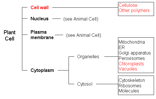
[c] Cells
[i] Every cell is covered by a membrane that controls what can enter and leave the cell. In all but quite primitive cells, a complex network of proteins provides organization and shape and, for animal cells, movement.
[ii] Within every cell are specialized parts for the transport of materials, energy transfer, protein building, waste disposal, information feedback, and even movement.
[iii] The work of the cell is carried out by the many different types of molecules it assembles, mostly proteins - long, usually folded chains made from 20 different kinds of amino-acid molecules. The function of each protein molecule depends on its specific sequence of amino acids and the shape the chain takes is a consequence of attractions between the chain's parts.
[iv] Before a cell divides, the instructions are duplicated so that each of the two new cells gets all the necessary information for carrying on.
[v] Complex interactions among the different kinds of molecules in the cell cause distinct cycles of activities, such as growth and division.
[vi] Gene mutations in a cell can result in uncontrolled cell division, called cancer. Exposure of cells to certain chemicals and radiation increases mutations and thus increases the chance of cancer.
[vii] A living cell is composed of a small number of chemical elements mainly carbon, hydrogen, nitrogen, oxygen, phosphorous, and sulfur.
[d] Interdependence of Life
[i] Ecosystems can be reasonably stable over hundreds or thousands of years. As any population of organisms grows, it is held in check by one or more environmental factors: depletion of food or nesting sites, increased loss to increased numbers of predators, or parasites.
[ii] Ecosystems tend to have cyclic fluctuations around a state of rough equilibrium. Human activities can, deliberately or inadvertently, alter the equilibrium in ecosystems.
[e] Flow of Matter and Energy
[i] At times, environmental conditions are such that plants and marine organisms grow faster than decomposers can recycle them back to the environment. Layers of energy-rich organic material have been gradually turned into great coal beds and oil pools by the pressure of the overlying earth.
[ii] By burning these fossil fuels, people are passing most of the stored energy back into the environment as heat and releasing large amounts of carbon dioxide.
[iii] The amount of life any environment can support is limited by the available energy, water, oxygen, and minerals, and by the ability of ecosystems to recycle the residue of dead organic materials.
[iv] The chemical elements that make up the molecules of living things pass through food webs and are combined and recombined in different ways. At each link in a food web, some energy is stored in newly made structures but much is dissipated into the environment as heat. Continual input of energy from sunlight keeps the process going.
[f] Evolution of Life
[i] The earth's present-day species developed from earlier, distinctly different species.
[ii] Molecular evidence substantiates the anatomical evidence for evolution and provides additional detail about the sequence in which various lines of descent branched off from one another.
[iii] Natural selection provides the following mechanism for evolution: Some variation in heritable characteristics exists within every species, some of these characteristics give individuals an advantage over others in surviving and reproducing, and the advantaged offspring, in turn, are more likely than others to survive and reproduce.
[iv] Heritable characteristics can be observed at molecular and whole-organism levels-in structure, chemistry, or behavior. These characteristics strongly influence what capabilities an organism will have and how it will react, and therefore influence how likely it is to survive and reproduce.
[v] New heritable characteristics can result from new combinations of existing genes or from mutations of genes in reproductive cells. Changes in other cells of an organism cannot be passed on to the next generation.
[vi] Natural selection leads to organisms that are well suited for survival in particular environments.
[vii] The theory of natural selection provides a scientific explanation for the history of life on earth as depicted in the fossil record and in the similarities evident within the diversity of existing organisms.
[viii] Life on earth is thought to have begun as simple, one-celled organisms, but once cells with nuclei developed, increasingly complex multicellular organisms evolved.
[ix] Evolution does not necessitate long-term progress in some set direction. Some evolutionary changes survive, many die out altogether, sometimes giving rise to more complex organisms.
|
1.1 - Cell Theory
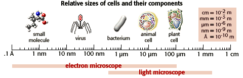
The cell is considered to be the smallest structure in biology that has all the properties of living things and an understanding of cells and the basics of cell structure and function is critical to making sense out of biology. Because of the limitations of the human eye, much of the early biological research concentrated on developing tools to help us see very small things. As imaging technology became more sophisticated, biological discoveries abounded.
To put things in perspective, the human eye has a resolution of about 100 µm. According to the chart below the plant cell is barely within our resolution but the components of the cell are best viewed with a light microscope..
In 1838, Theodor Schwann and Matthias Schleiden were talking about their studies on cells. When Schwann heard Schleiden describe plant cells with nuclei, he was struck by the similarity of these plant cells to cells he had observed in animal tissues. The two scientists went immediately to Schwann's lab to look at his slides. Schwann published his book on animal and plant cells the next year. When the cell theory was proposed in 1838, cell biology research was forever changed. The cell theory states that:
[1] All life forms are made from one or more cells.
[2] Cells only arise from pre-existing cells.
[3] The cell is the smallest form of life.
These statements seem self evident nowadays, but this theory actually only dates from the mid 19th century. Before the mid 19th century many people believed in spontaneous generation. This is the idea that living things can develop from non-living things. For example, it was widely believed that flies actually developed from rotten meat or that bacteria developed from stagnant water, or that frogs developed from the mud at the bottom of ponds, or that a horse hair put in water would turn into a worm. Scientists cleared up these beliefs and now we accept the notion that under current conditions, life does not arise from non life.
1.2 - Prokaryotic Cells
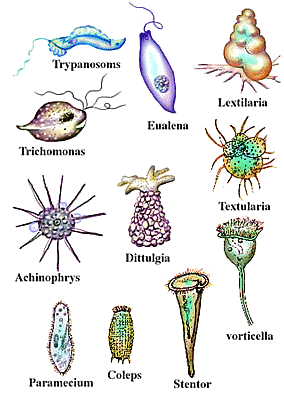
Prokaryotes are single-celled organisms that lack a nucleus and that do not have their genetic material organized into chromosomes. They constitute the Kingdom Monera, Which is split into two divisions: the Schizophyta ( traditionally called bacteria ) and the Cyanophyta (formerly known as blue-green algae ). Here are some examples of prokaryotic cells...
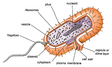
The surface structure includes a capsule; a layer of polysaccharide (sometimes proteins) that protects the bacterial cell and is often associated with pathogenic bacteria because it serves as a barrier against phagocytosis by white blood cells. Moving inwards, the cell wall is composed of peptidoglycan (polysaccharides + protein), the cell wall maintains the overall shape of a bacterial cell. The three primary shapes in bacteria are coccus (spherical), bacillus (rod-shaped) and spirillum (spiral). Mycoplasma are bacteria that have no cell wall and therefore have no definite shape. The plasma membrane is a lipid bilayer much like the cytoplasmic (plasma) membrane of other cells. There are numerous proteins moving within or upon this layer that are primarily responsible for transport of ions, nutrients and waste across the membrane.
Bacteria may have appendages such as pili and flagella. Pili are are hollow, hairlike structures made of protein allow bacteria to attach to other cells. A specialized pilus, the sex pilus, allows the transfer from one bacterial cell to another. Pili (sing., pilus) are also called fimbriae (sing., fimbria). The purpose of flagella (sing., flagellum) is motility. Flagella are long appendages which rotate by means of a "motor" located just under the cytoplasmic membrane. Bacteria may have one, a few, or many flagella in different positions on the cell.
1.3 - Eukaryotic Cells
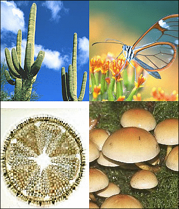
Eukaryotic cells (from the Greek meaning truly nuclear) comprise all of the life kingdoms except monera. They can be easily distinguished through a membrane-bound nucleus. All species in the Eukaryota domain (protists, fungi, plants, and animals) have eukaryotic cells. Individual protists are small and have only one cell, while individual plants and animals can have trillions of cells. Complex creatures like humans have special cells for particular functions such as carrying oxygen around the body, digesting food, or making bone.
Clockwise from upper left: plantae, animalia, fungi, and protist.
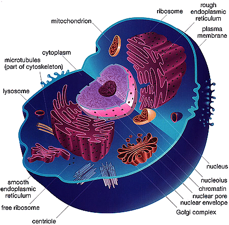
The eukaryotic cell contains organelles, which are defined as membrane-bound structures such as nucleus, mitochondria, chloroplasts, endoplasmic reticulum (ER), Golgi apparatus, lysosomes, vacuoles, peroxisomes, etc. Let's take a look at some models of plant and animal cells,
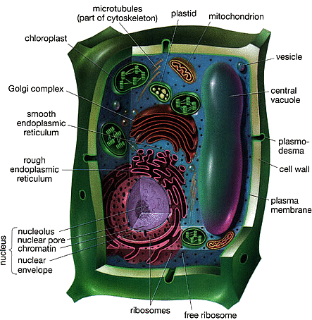
differences [plant vs. animal cells] are color coded red
differences [plant vs. animal cells] are color coded red
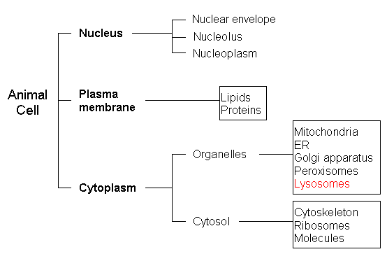
The nucleus is the most conspicuous organelle found in a eukaryotic cell. It houses the cell's chromosomes and is the place where almost all DNA replication and RNA synthesis occur. The nucleus is spheroid in shape and separated from the cytoplasm by a membrane called the nuclear envelope. The nuclear envelope isolates and protects a cell's DNA from various molecules that could accidentally damage its structure or interfere with its processing. During processing, DNA is transcribed, or synthesized, into a special RNA, called mRNA. This mRNA is then transported out of the nucleus, where it is translated into a specific protein molecule. In prokaryotes, DNA processing takes place in the cytoplasm.
Ribosomes are found in both prokaryotes and eukaryotes. The ribosome is a large complex composed of many molecules, including RNAs and proteins, and is responsible for processing the genetic instructions carried by an mRNA. The process of converting an mRNA's genetic code into the exact sequence of amino acids that make up a protein is called translation. Protein synthesis is extremely important to all cells, and therefore a large number of ribosomessometimes hundreds or even thousandscan be found throughout a cell. Ribosomes float freely in the cytoplasm or sometimes bind to another organelle called the endoplasmic reticulum. Ribosomes are composed of one large and one small subunit, each having a different function during protein synthesis.
Mitochondria are self-replicating organelles that occur in various numbers, shapes, and sizes in the cytoplasm of all eukaryotic cells. As mentioned earlier, mitochondria contain their own genome that is separate and distinct from the nuclear genome of a cell. Mitochondria have two functionally distinct membrane systems separated by a space: the outer membrane, which surrounds the whole organelle; and the inner membrane, which is thrown into folds or shelves that project inward. These inward folds are called cristae. The number and shape of cristae in mitochondria differ, depending on the tissue and organism in which they are found, and serve to increase the surface area of the membrane.
Mitochondria play a critical role in generating energy in the eukaryotic cell, and this process involves a number of complex pathways. Let's break down each of these steps so that you can better understand how food and nutrients are turned into energy packets and water. Some of the best energy-supplying foods that we eat contain complex sugars. These complex sugars can be broken down into a less chemically complex sugar molecule called glucose. Glucose can then enter the cell through special molecules found in the membrane, called glucose transporters. Once inside the cell, glucose is broken down to make adenosine triphosphate (ATP), a form of energy
Chloroplasts are similar to mitochondria but are found only in plants. Both organelles are surrounded by a double membrane with an intermembrane space; both have their own DNA and are involved in energy metabolism; and both have reticulations, or many foldings, filling their inner spaces. Chloroplasts convert light energy from the sun into ATP through a process called photosynthesis.
The endoplasmic reticulum (ER) is the transport network for molecules targeted for certain modifications and specific destinations, as compared to molecules that will float freely in the cytoplasm. The ER has two forms: the rough ER and the smooth ER. The rough ER is labeled as such because it has ribosomes adhering to its outer surface, whereas the smooth ER does not. Translation of the mRNA for those proteins that will either stay in the ER or be exported (moved out of the cell) occurs at the ribosomes attached to the rough ER. The smooth ER serves as the recipient for those proteins synthesized in the rough ER. Proteins to be exported are passed to the Golgi apparatus, sometimes called a Golgi body or Golgi complex, for further processing, packaging, and transport to a variety of other cellular locations.
Lysosomes and peroxisomes are often referred to as the garbage disposal system of a cell. Both organelles are somewhat spherical, bound by a single membrane, and rich in digestive enzymes, naturally occurring proteins that speed up biochemical processes. For example, lysosomes can contain more than three dozen enzymes for degrading proteins, nucleic acids, and certain sugars called polysaccharides. One function of a lysosome is to digest foreign bacteria that invade a cell. Other functions include helping to recycle receptor proteins and other membrane components and degrading worn out organelles such as mitochondria. Lysosomes can even help repair damage to the plasma membrane by serving as a membrane patch, sealing the wound.
1.4 - Cell Membranes
According to the old adage, oil and water don't mix. As simple as it sounds, this principle is the construction secret behind one of evolution's most successful structures: the cell membrane.
While the plant cell has a rigid cell wall, an animal cell membrane is a flexible lipid bilayer. Lipids are the one class of large biological molecules that does not include polymers. They are grouped together because they share one important chemical property: they have little or no affinity for water. The hydrophobic behavior of lipids is based on their molecular structure. The lipid molecules (mostly phospholipids) that make up the membrane have a polar, hydrophilic head and two hydrophobic hydrocarbon tails. When the lipids are immersed in an aqueous solution the lipids spontaneously bury the tails together and leave the hydrophilic heads exposed. Thus this is a handy membrane to use, because it can automatically fix itself when torn. There are three different major classes of lipid molecules - phospholipids, cholesterol, and glycolipids. Different membranes have different ratios of the three lipids.
Symbol, structural formula and space-filling model
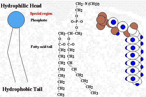
Phospholipids are described as being amphipathic, having both a hydrophobic and a hydrophilic region. Their tails, which consist of hydrocarbons, are hydrophobic and are excluded from water. Their heads, however, which consist of the phosphate group and its attachments, are hydrophilic, and have an affinity for water. Because of their structure, when phospholipids are added to water, they self-assemble into aggregates so that the phosphate heads make contact with the water and the hydrophobic hydrocarbon tails are restricted to water-free areas.
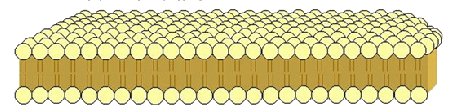
Membranes are not static sheets of molecules locked rigidly in place. A membrane is held together primarily by hydrophobic interactions, which are much weaker than covalent bonds. Most lipids are randomly mobile in the plane of the membrane with an average migration rate of 22 μm (micrometers) per second. It is rare, however, for a molecule to flip-flop transversely across the membrane, switching from one phospholipid layer to the other; to do so, the hydrophilic part of the molecule would have to cross the hydrophobic core of the membrane.
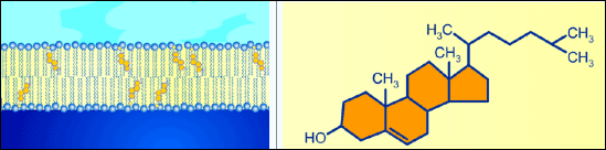
The steroid cholesterol, which is wedged between phospholipid molecules in the plasma membranes of animals, helps stabilize the membrane. At relatively warm temperatures, for example, 370C, the body temperature of humans, cholesterol makes the membrane less fluid by restraining the movement of phospholipids. However, because cholesterol hinders the close packing of phospholipids, it also lowers the temperature required for the membrane to solidify.
among phospholipid tails in the bilayer
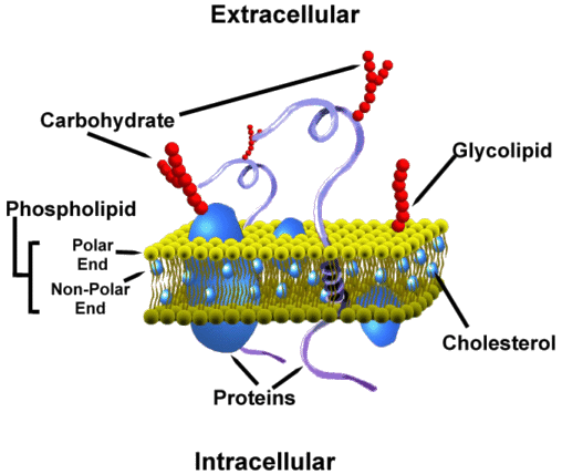
According to the accepted current theory, known as the fluid mosaic model, the biological membrane is a collage of many different proteins embedded in the fluid matrix of the lipid bilayer. The lipid bilayer is the main fabric of the membrane, and its structure creates a semi-permeable membrane. Proteins are the most structurally sophisticated molecules known, and account for more than 50% of the dry weight of most cells. Although they are diverse, humans have tens of thousands of different proteins, each with a specific structure and function; they are all polymers constructed from the same set of 20 amino acids. Membrane proteins are classified into two major categories, Integral proteins and Peripheral proteins. Integral proteins are generally transmembrane proteins, with hydrophobic regions that completely span the hydrophobic interior of the membrane. The hydrophilic ends of the molecule are exposed to the aqueous solutions on either side of the membrane. Proteins are much larger than lipids and move more slowly, but some do drift. Some membrane proteins seem to move in a highly directed manner, however, many others seem to be held virtually immobile by their attachment to the cytoskeleton. Peripheral proteins are not embedded in the lipid bilayer at all; they are loosely bound to the surface of the membrane, often to the exposed parts of integral proteins. Glycoproteins and glycolipids have carbohydrates attached to their extracellular domains.
1.5 - Cell Division
For most unicellular organisms, reproduction is a simple matter of cell duplication, also known as replication. But for multicellular organisms, cell replication and reproduction are two separate processes. Multicellular organisms replace damaged or worn out cells through a replication process called mitosis, the division of a eukaryotic cell nucleus to produce two identical daughter nuclei.
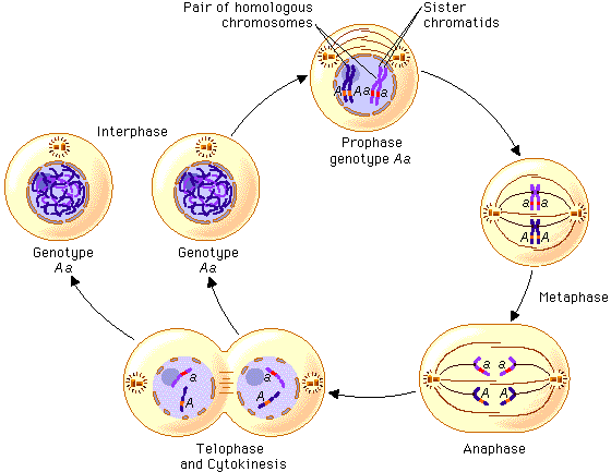
Mitosis is the process by which the diploid nucleus (having two sets of homologous chromosomes) of a somatic cell divides to produce two daughter nuclei, both of which are still diploid. Before a cell enters mitosis, we say the cell is in interphase, the state of a eukaryotic cell when not undergoing division. Every time a cell divides, it must first replicate all of its DNA. Because chromosomes are simply DNA wrapped around protein, the cell replicates its chromosomes also. These two chromosomes, positioned side by side, are called sister chromatids and are identical copies of one another. Before this cell can divide, it must separate these sister chromatids from one another. To do this, the chromosomes have to condense. This stage of mitosis is called prophase. Next, the nuclear envelope breaks down, and a large protein network, called the spindle, attaches to each sister chromatid. The chromosomes are now aligned perpendicular to the spindle in a process called metaphase. Next, "molecular motors" pull the chromosomes away from the metaphase plate to the spindle poles of the cell. This is called anaphase. Once this process is completed, the cells divide, the nuclear envelope reforms, and the chromosomes relax and decondense during telophase. The cell can now replicate its DNA again during interphase and go through mitosis once more.
To reproduce, eukaryotes must first create special cells called gameteseggs and spermthat then fuse to form the beginning of a new organism. Gametes are but one of the many unique cell types that multicellular organisms need to function as a complete organism.
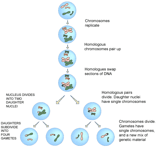
Meiosis, a type of nuclear division, occurs only in reproductive cells and results in a diploid cell (having two sets of chromosomes) giving rise to four haploid cells (having a single set of chromosomes). Each haploid cell can subsequently fuse with a gamete of the opposite sex during sexual reproduction.
Meiosis I refers to the first of the two divisions and is often called the reduction division. This is because it is here that the chromosome complement is reduced from diploid (two copies) to haploid (one copy). Interphase in meiosis is identical to interphase in mitosis. At this stage, there is no way to determine what type of division the cell will undergo when it divides. Meiotic division will only occur in cells associated with male or female sex organs. Prophase I is virtually identical to prophase in mitosis, involving the appearance of the chromosomes, the development of the spindle apparatus, and the breakdown of the nuclear membrane. Metaphase I is where the critical difference occurs between meiosis and mitosis. In mitosis, all of the chromosomes line up on the metaphase plate in no particular order. In Metaphase I, the chromosome pairs are aligned on either side of the metaphase plate. It is during this alignment that the chromatid arms may overlap and temporarily fuse, resulting in what is called crossovers. During Anaphase I, the spindle fibers contract, pulling the homologous pairs away from each other and toward each pole of the cell. In Telophase I, a cleavage furrow typically forms, followed by cytokinesis, the changes that occur in the cytoplasm of a cell during nuclear division; but the nuclear membrane is usually not reformed, and the chromosomes do not disappear. At the end of Telophase I, each daughter cell has a single set of chromosomes, half the total number in the original cell, that is, while the original cell was diploid; the daughter cells are now haploid.
Meiosis II is quite simply a mitotic division of each of the haploid cells produced in Meiosis I. There is no Interphase between Meiosis I and Meiosis II, and the latter begins with Prophase II. At this stage, a new set of spindle fibers forms and the chromosomes begin to move toward the equator of the cell. During Metaphase II, all of the chromosomes in the two cells align with the metaphase plate. In Anaphase II, the centromeres split, and the spindle fibers shorten, drawing the chromosomes toward each pole of the cell. In Telophase II, a cleavage furrow develops, followed by cytokinesis and the formation of the nuclear membrane. The chromosomes begin to fade and are replaced by the granular chromatin, a characteristic of interphase. When Meiosis II is complete, there will be a total of four daughter cells, each with half the total number of chromosomes as the original cell. In the case of male structures, all four cells will eventually develop into sperm cells. In the case of the female life cycles in higher organisms, three of the cells will typically abort, leaving a single cell to develop into an egg cell, which is much larger than a sperm cell.
Most unicellular organisms (such as bacteria) create their next generation by replicating all of their parts and then splitting into two cells, a type of asexual reproduction called binary fission. This process spawns not just two new cells, but also two new organisms. As just noted, this type of asexual reproduction theoretically results in two identical cells. However, bacterial DNA has a relatively high mutation rate. This rapid rate of genetic change is what makes bacteria capable of developing resistance to antibiotics and helps them exploit invasion into a wide range of environments.
Similar to more complex organisms, bacteria also have mechanisms for exchanging genetic material. Although not equivalent to sexual reproduction, the end result is that a bacterium contains a combination of traits from two different parental cells. Three different modes of exchange have thus far been identified in bacteria. Conjunction involves the direct joining of two bacteria, which allows their circular DNAs to undergo recombination. Bacteria can also undergo transformation by absorbing remnants of DNA from dead bacteria and integrating these fragments into their own DNA. Lastly, bacteria can exchange genetic material through a process called transduction, in which genes are transported into and out of the cell by bacterial viruses, called bacteriophages, or by plasmids, an autonomous self-replicating extrachromosomal circular DNA.
|
2.1 - Chemical elements and water
...
|
3.1 - Chromosomes, genes, alleles, and mutations
...
|
4.1 - Tissues and Systems
...
|
5.1 - Evolution of Life
...
[1] A Beka, Living Creation, Work-Text, Pensacola, Florida.
[2] Dobell, C. Antony van Leeuwenhoek and His "Little Animals" (Dover, New York, 1960)
[3] Mayr, E. The Growth of the Biological Thought (Belknap, Cambridge, MA, 1982)
[4] Singer, S. A Short History of Biology (Clarendon, Oxford, 1931)